Low BMI in Axial Spondyloarthritis Patients Linked to Weaker Bones, Study Suggests
Written by |


After analyzing 80 axial spondyloarthritis (axSpA) patients, a French research team found a lower bone mineral density (BMD, or weaker bones) among those who also have a lower body mass index (BMI).
The results were reported in a study titled “Prevalence and risk factors of low bone mineral density in spondyloarthritis and prevalence of vertebral fractures,” published in the journal BMC Musculoskeletal Disorders.
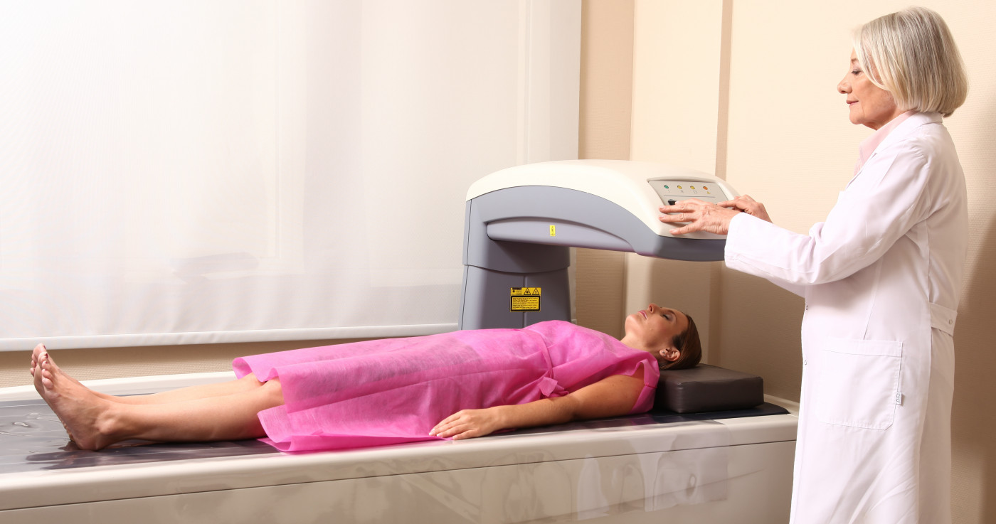
About 13% of axSpA patients have osteoporosis caused by bone loss. As a result, they routinely undergo a dual energy X-ray absorptiometry (DXA) examination, a low radiation standard technique for measuring BMD. Doctors usually take measurements from the femur and in the lumbar spine in the anterior-posterior projection.
In this study, researchers used a newer-generation densitometry machine where patients may be in a supine position (the body lying down with the face up). This allowed the team to take lateral spine BMD measurements. They also looked for vertebrae fractures and calcifications (syndesmophytes) by vertebral fracture assessment (VFA).
“The utility of our study is that it investigated DXA BMD in patients with axial SpA regardless of disease severity or duration, that it included lateral spine measurement, and that VFA was performed in all cases, enabling the detection of fractures and analysis of syndesmophytes,” researchers wrote.
Using this new approach, the team evaluated the risk factors of low BMD and vertebral fractures in 37 women and 52 men who met the clinical criteria for axial SpA. The group had an average age of 44 years and a mean disease duration of 10.2 years. In total, 48.3% of the patients were diagnosed with osteopenia, and 6.7% with osteoporosis.
Researchers found lower spine BMD values on lateral measurement than on anterior-posterior measurement (0.809 vs. 1) but only detected two additional cases of osteopenia. Also, by VFA analysis, a fracture was detected in five patients (6.25%), with a total of eight fractures detected.
They studied the different factors that could lower BMD, such as age, sex, lower BMI, vitamin D levels, and physician’s global assessment.
Researchers concluded that BMI was the strongest factor associated with low BMD.
“Our study found a high prevalence (around 50%) of low BMD in SpA. Conversely, the prevalence of osteoporosis (6.7% according to WHO criteria) and vertebral fractures (6.2%) was lower than generally reported in the literature,” researchers wrote.
Nonetheless, the team also suggested that less physical activity and post-menopause status could lead to lower BMD.
“A loss of mobility is also a classic risk factor for bone loss. It can be assumed that disease-related changes in functional capacity may be one of the causes of bone loss in SpA,” researchers wrote. “We didn’t take into account post-menopausal status that was not available and could interfere with women’s bone density.”
Researchers were unable to find any association between low BMD and smoking, the use of non-steroidal anti-inflammatory drugs (NSAIDs), or vitamin D supplements.